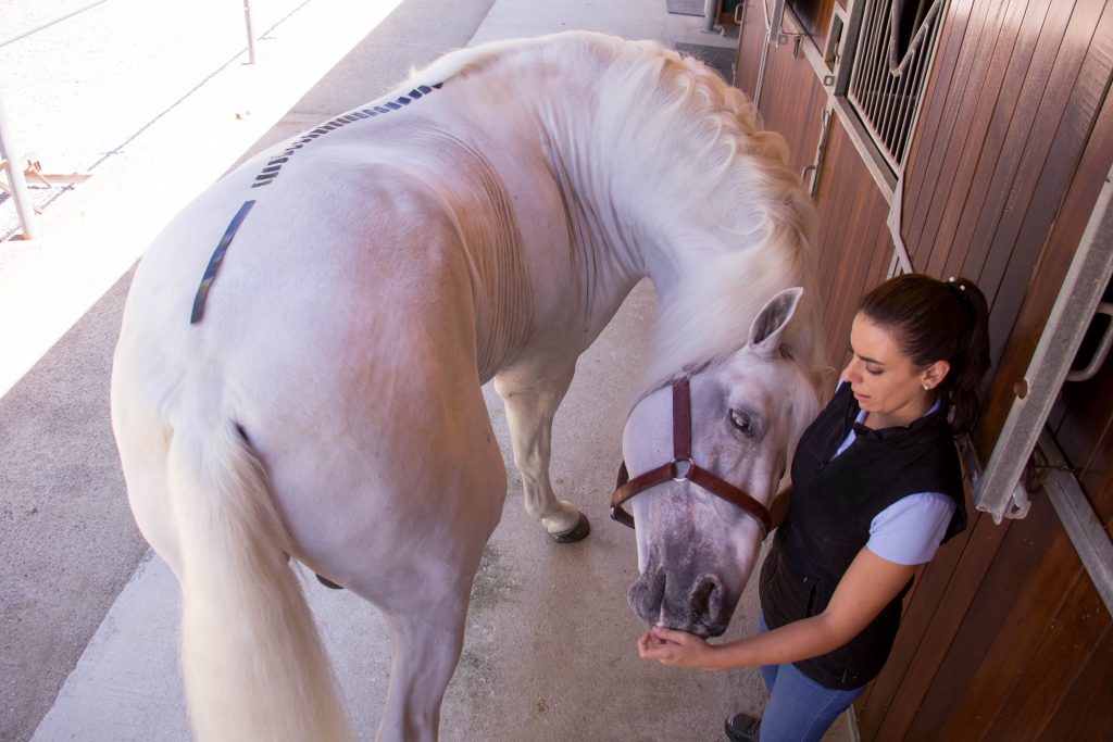
Any musculoskeletal alteration comes along with postural patterns compensations that seem to be constant and are very well defined in humans according to Myers T (ix) but not as well described in horses. Elbrond & Shultz (2014) describes Myofascial Kinetic Lines in horses. Below you can find a detailed comparison of the backline on equines based on a level III of scientific evidence.
SUPERFICIAL DORSAL LINE:
It begins at the posterior base of the third phalanx and connects the entire posterior aspect of the hind limbs and hindquarters, the back and the dorsal aspect of the horse’s neck, ending in the skull. It supports the horse’s body in extension avoiding its hang due to:
- The weight of the neck and head
- The weight of the chest and abdomen
- The tension of the frontal line
These postural functions require an increase in fibers type I and require very dense and resistant fasciae. Specifically, postural patterns compensation appear to be:
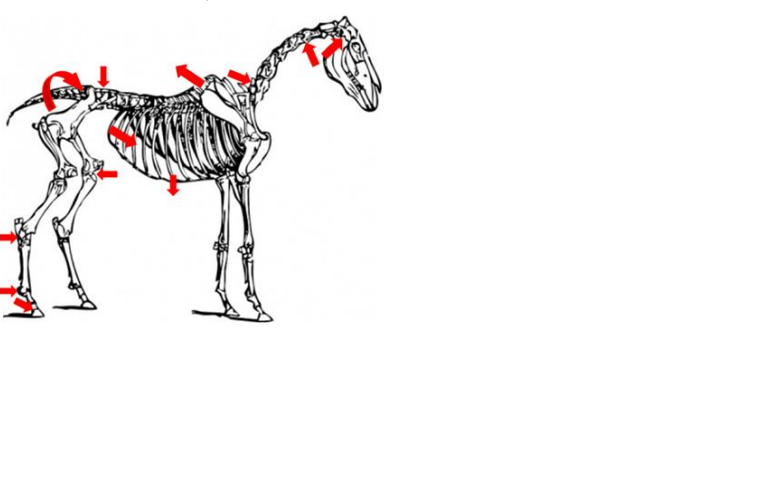
- Tendency to verticalization of the third phalanx, the tendon of the deep digital flexor muscle can be found in a short position and the tendon of the long digital extensor muscle in a long position
- Tendency to flexure the fetlock so there is a limitation of the hyperextension on the fetlock.
- Tendency to hock extension due to possible short position of the deep digital flexor muscle and the gastrocnemius muscle. The longus extensor digitorum muscle can be found in a long position.
- A tendency to extend the stifle as the semimembranosus and quadriceps femoris muscles may be in a short position while the biceps femoris muscle and the semitendinosus muscle are in a long position.
- Anterior translation and anteversion of the pelvis due to possible short position of the gluteus medius, deep gluteus and piriformis muscles. The gluteus superficialis and tensor fasciae lata muscles may be in a long position.
- Tendency to nutation of the sacrum. The short position of the biceps femoris and the multifidus muscle in its lumbo-sacral portion can produce this alteration.
- Tendency to lumbar lordosis due to block in short position of the multifidus muscle and the psoas minor muscle.
- Tendency to horizontalization of the withers due to the possible blocking in short position of the lumbar longissimus and thorax longissimus muscles.
- Tendency to anterior translation and tilt of the rib cage due to possible blockage in short position of the cranial serratus dorsalis muscle, the external intercostal muscles, the ribs levator and the rectus of the abdominal. The muscle serratus dorsalis caudal, the internal intercostal muscles, and the transverse muscle of the abdominal may be in a long position.
- Tendency to increase low cervical lordosis as the cervical iliocostal muscle, the longissimus of the neck and the multifidus muscle in its cervical portion may be in a short position.
- Tendency to rectify high cervical kyphosis and to extend the occipital-atlas-axis complex. The blockade in short position of the semispinal muscle of the head, the longus muscle of the head and the longus muscle of the atlas together with the multifidus muscle in its cervical portion can produce this alteration. In addition, it is possible to observe a tendency to the anterior translation of the head and an anterior sliding of the occiput with respect to the atlas.
ix Thomas W. Myers